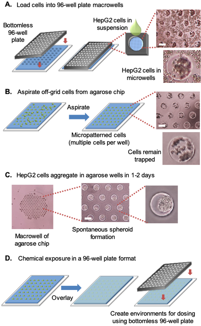
Figure 2.

HepG2 cells aggregate once loaded into agarose microwells. The intact spheroids can be handled on the agarose chip in a standard 96-well array. (A) HepG2 cells fall into the agarose microwells by gravity. (B) Excess cells are removed from the agarose chip by aspirating between macrowells. (C) HepG2 spheroids form spontaneously in microwells. (D) Once spheroids have formed, they can be immobilized on the chip with an agarose overlay. A bottomless 96-well plate is then placed over the chip, creating distinct environments for chemical exposure. White scale bar, 250 μm. Note that each microwell is ∼250 μm in diameter.
