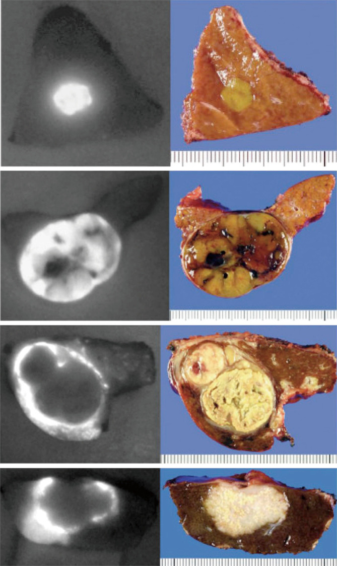
Figure 1.

Differential patterns of fluorescence signals on the cut surface of different liver cancers using ICG. ICG administered pre-operatively gives a fluorescence signal over the liver when imaged with a NIR-fluorescence camera. The patterns can be classified into bright homogenous “total” fluorescence type (A) well-differentiated hepatocellular carcinoma (HCC), partial fluorescence type (B) moderately differentiated HCC, and rim-type fluorescence (C), poorly differentiated HCC (upper) and CLM (lower). Adapted from (104). ICG, indocyanine green; CLM, colorectal liver metastases.
