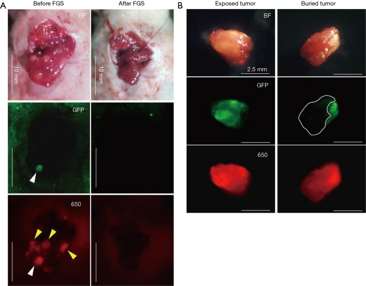
Figure 3.
Visualizing a liver metastasis in an orthotopic model of colon cancer with a fluorescent antibody. Comparison between visible wavelength fluorescence and near-infrared fluorescence signal using anti-CEA antibody conjugated to DyLight650 visualized using a Mini Maglite® LED Pro flash light (Mag Instrument) with an excitation filter (ET640/30X, Chroma Technology Corporation) and a Canon EOS 60D digital camera with an EF-S18-55 IS lens (Canon) and an emission filter (HQ700/75M-HCAR, Chroma Technology Corporation). (A) The surface-exposed tumor (white arrow head) was clearly detected under both GFP and anti-CEA-DyLight650 navigation. In contrast, the tumors covered with normal tissues (yellow arrow heads) were detected only under anti-CEA-DyLight650 navigation. No residual tumor was detected after FGS. (B) Representative gross images of excised tumors (left panel: exposed tumor, right panel: buried tumor). Upper panels indicate bright field (BF) images; middle and lower panels indicate fluorescence images for GFP and DyLight 650 [650], respectively. The area surrounded by a white broken line indicates the buried part of the excised tumor. CEA650 fluorescence was able to penetrate normal liver tissue and visualize the buried part of the tumor, which was not detected by GFP fluorescence. Scale bars: 10 mm (A) and 2.5 mm (B). Adapted from (119). CEA, carcinoembryonic antigen; GFP, green fluorescence protein.

