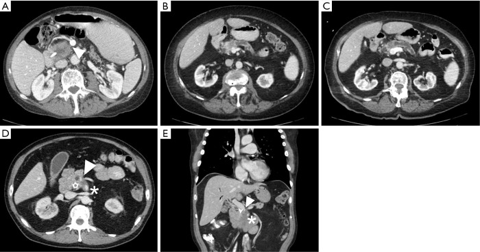
Figure 1.
CT images of resectable, borderline resectable before and after treatment with neoadjuvant therapy and locally advanced pancreatic cancers. (A) Resectable pancreatic head mass with a clear fat plane (arrow) separating tumor from the superior mesenteric vein; (B) borderline resectable pancreatic head mass with tumor in contact with the superior mesenteric vein with slight distortion of the vessel. Note the distal pancreatic duct appears dilated; (C) borderline resectable pancreatic head mass following neoadjuvant chemotherapy with continued abutment and a distortion of a portion of the superior mesenteric vein; (D,E) locally advanced pancreatic head mass with encasement of the superior mesenteric vein (arrow) and superior mesenteric artery (star).

