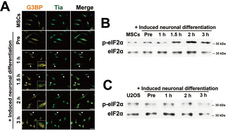
Fig. 1. SGs aggregation during neuronal differentiation.
(A) Human bone marrow-mesenchymal stem cells (hBM-MSCs) were incubated with pre-induction medium for one day and exposed to neuronal induction medium for 0, 1, 1.5, 2, and 3 h prior to fixation and immunocytochemical staining for G3BP (orange) and TIA-1 (green). SG-positive cells are indicated by arrowheads. Scale bars = 50 μm. (B) The protein expression of eIF2α and p-eIF2α was measured by immunoblot analysis after hBM-MSCs were incubated with neuronal induction medium for the indicated time. (C) U2OS cells were incubated with differentiation medium as shown in panel A. Total protein from induced U2OS cells was measured by immunoblot analysis using antibodies for eIF2α and p-eIF2α. “Pre,” indicates hBM-MSCs incubated with pre-induction medium for one day.

