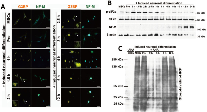
Fig. 2. SGs are associated with neuronal differentiation of stem cells.
(A) hBM-MSCs were treated with neuronal induction medium for 0, 1, 1.5, 2, 2.5, 3, 4, 6, and 12 h. Cells were fixed and immunostained for G3BP (orange) and NF-M (green). Arrowheads indicate SG-positive cells. Scale bars = 50 μm. (B) hBM-MSCs were induced to differentiate into neurons. Total proteins were extracted from the differentiating cells at the indicated times and p-eIF2α and NF-M expression levels were measured by immunoblot analysis. (C) The cells were labeled with 25 μM AHA for 4 h and harvested at the indicated times. Newly synthesized proteins were measured by immunoblot analysis for biotin-azide and streptavidin conjugation. “–AHA” indicates not labeled with AHA.

