. 2020 Oct 26;12(5):299–306. doi: 10.4047/jap.2020.12.5.299

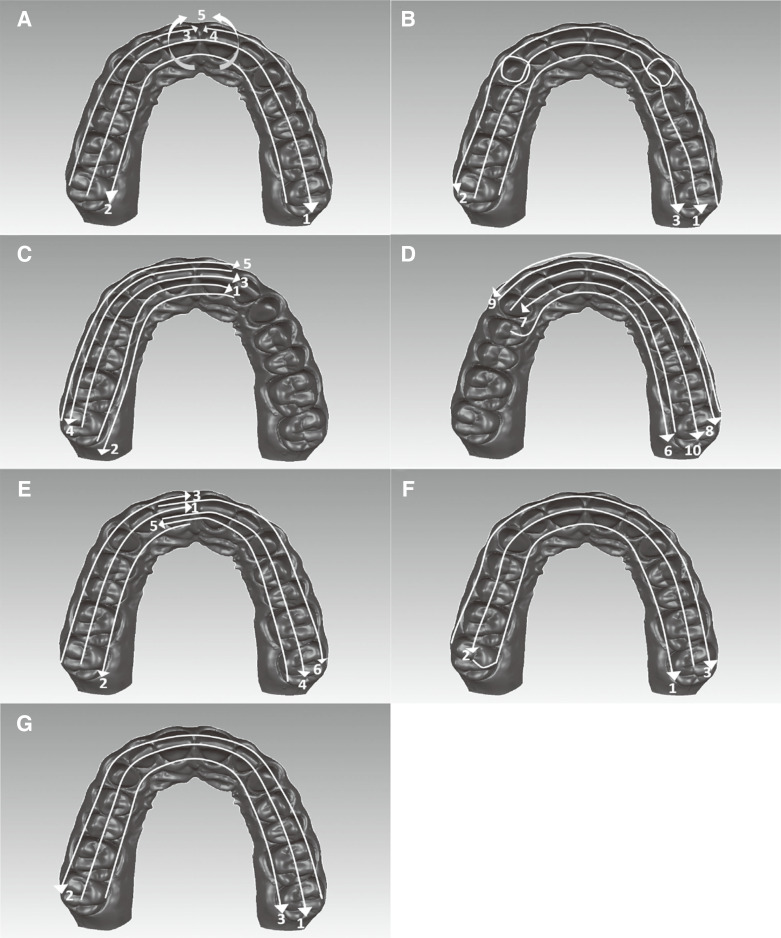
Fig. 1. Scan strategy for the ScanR. (A) iTero Element 2. The scan was started from the occlusal side of the right molars to the left occlusal side (1), continued the palatal side in the reverse direction (2), and toward the buccal side to the midline (3), moved to the opposite side and scanned to the midline (4), scanned by rolling over the anterior teeth (5). (B) Trios 3. Started from the occlusal side of the right molars to the left occlusal side by rolling over palatal, buccal, distal, and mesial side of preparations (1), continued the buccal side in the reverse direction (2), and toward the palatal side (3) (C) Omnicam, first half of arch. Started from the occlusal side of the right molars and tilted the scanner by 45° in palatal direction (1), continued the palatal side (90°) from the left lateral to the right molar (2), toward the occlusal side in the reverse direction (3), scanned the buccal side with 45° tilt (4) and 90° tilt (5). (D) Omnicam, second half of arch. Started from the occlusal side of the right premolar and continued the palatal side with 90° (6) and 45° (7) tilt, followed the buccal side with 45° (8) and 90° (9) tilt, and finally occlusal side (10). (E) Planmeca Emerald. Started from occlusal side of the right molar to the midline (1), continued the palatal side (2), and buccal side (3), scanned the second half similar to the first half, starting from the central tooth. (F) Primescan. Scanned the palatal side (1), occlusal side (2), and buccal side (3), respectively. (G) Virtuo Vivo. Scanned the occlusal side (1), buccal side (2), and palatal side (3), respectively (not defined a specific scan strategy by manufacturer).

Fig. 1