Figure 7.
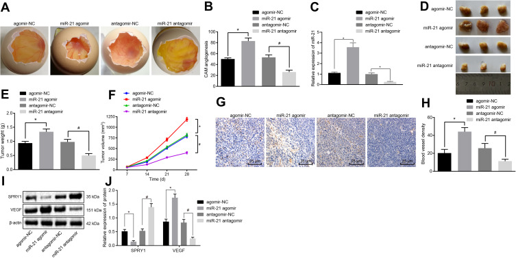
The elevation of miR-21 promotes the angiogenesis in vivo. CAM model and nude mice were transduced with the miR-21 agomir, agomir-NC, miR-21 antagomir or antagomir-NC. (A) The angiogenesis observed in CAM model (scale bar = 2 mm; n = 8). (B) The branching of blood vessels embedded in Matrigel in the surrounding 5 mm area counted after 10-d incubation in CAM model (n = 10). (C) The expression pattern of miR-21 in the serum of CAM model as determined using RT-qPCR. (D–F) The formation, volume, and weight of tumor in nude mice xenografted with KYSE-150 cells (n = 8). (G) MVD evaluation of mice tumor tissues (scale bar = 25 μm; n = 8). (H) MVD evaluation of mice tumor tissues (n = 8). (I, J) The protein expression patterns of SPRY1 and VEGF determined using immunoblotting (SPRY1: 35 kDa, VEGF: 151 kDa) (n = 8). *p < 0.05 vs treatment with agomir-NC, #p < 0.05 vs treatment with antagomir-NC. Comparisons among multiple groups were conducted by one-way ANOVA, followed by a Tukey’s post hoc test. Statistical analysis in relation to time-based measurements within each group was realized using repeated measures ANOVA, followed by a Bonferroni’s post hoc test (E).

