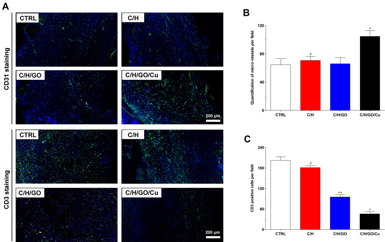
Figure 8.
The effect of GO/Cu nanocomposite-modified dressings on inflammatory infiltration and angiogenesis in regenerated tissues.
Notes: (A) Representative images of immunofluorescent CD31 and CD3 staining of wound sections in the CTRL, C/H, C/H/GO and C/H/GO/Cu groups at the day of sacrifice. Endothelial cells stained with CD31 antibody and inflammatory cells stained with CD3 antibody are fluorescent green, whereas cell nuclei stained with DAPI are fluorescent blue. (B) Quantification of microvessels per randomly selected microscopic field in various groups. (C) Quantification of CD3-positive cells per randomly selected microscopic field in various groups. *Represents a significant difference compared with the other groups (p<0.01). **Represents a significant difference compared with the CTRL and C/H groups (p<0.01). #Represents a significant difference compared with the CTRL group (p<0.05).

