Fig. 3.
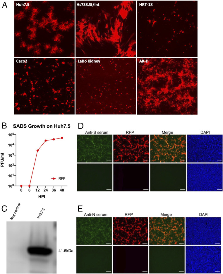
Host range of SADS tRFP. (A) Cultures of human Huh7.5 liver, human Hs738.St/Int stomach-intestine, human colo-rectal HRT-18 tumor, Caco2, LaBo kidney, and feline AK-D lung cells were infected with SADS tRFP. All cell types, with the exception of Caco2 and Huh7.5 cells, were cultured in the presence of trypsin to enhance virus infectivity. Cultures were visualized at 48 hpi for fluorescence at 10× magnification. (B) Growth of SADS-tRFP in Huh 7.5 cells. (C) Cultures of Huh7.5 cells were infected with SADS-CoV and lysed for analysis by Western blot. N protein has a molecular mass of ∼41.6 kDa. Immunofluorescent images of Vero CCL-81 cells infected with mock or SADS-CoV tRFP virus and stained with mouse anti-N (D) or anti-S (E) sera. Cultures were fixed at 24 hpi and viral proteins were visualized by immunostaining with antisera isolated from VRP S or VRP N vaccinated mice. Scale bars represent 100 µm.

