Fig. 4.
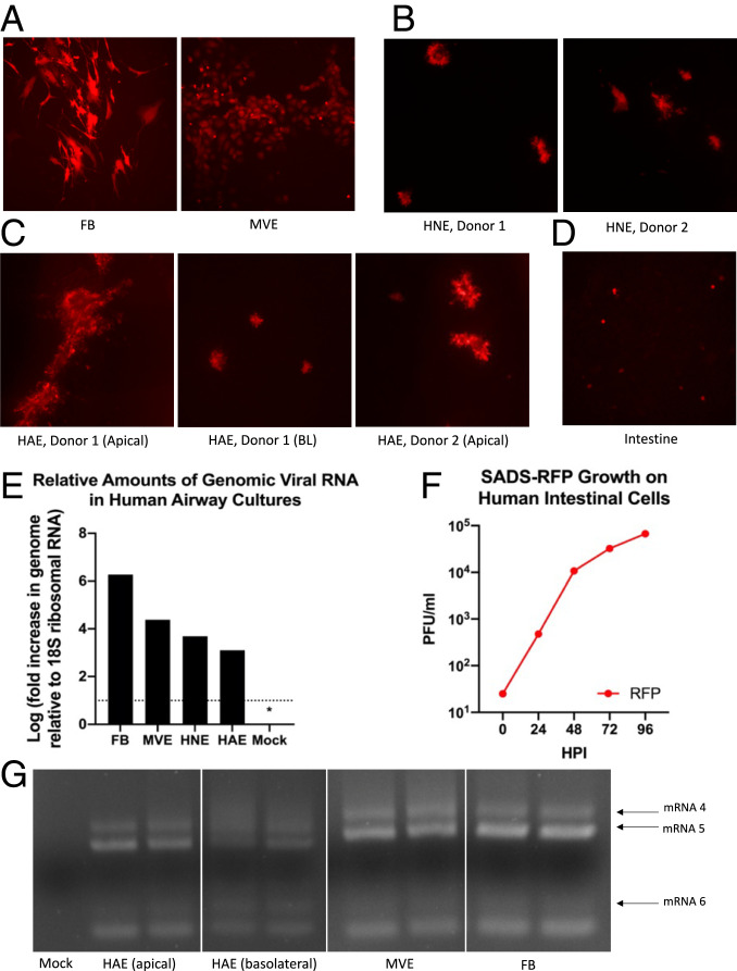
Susceptibility of human primary cells to SADS-CoV. (A) Human FB and MVE cells (n = 3, each) were infected with SADS-CoV. By 72 hpi, abundant infection of both FB and MVE cells by SADS-RFP was observed. (B) HNE cells from two donors (n = 3) were infected with SADS-RFP, demonstrating comparable RFP infection by 72 hpi. (C) HAE cells were infected both basolaterally and/or apically (donor 1) or apically (donor 2) and observed for 96 h or 72 h, respectively. (D) Human primary intestinal cultures (n = 2) were infected with SADS-RFP and were observed for 96 hpi. All fluorescent images were taken at 10× magnification. (E) qRT-PCR of genomic mRNA from primary human lung cells infected with SADS-CoV. Cultures from various codes were averaged to determine amounts of genomic viral RNA. No detectable viral signal was observed in mock-infected cultures from each cell type, as indicated by the * representing half the limit of detection. Levels of viral genome were determined in infected and mock cultures, relative to 18S ribosomal RNA. (F) Virus samples were taken from primary intestinal cells every 24 hpi, and growth determined by plaque assay. (G) RT-PCR of leader containing transcripts, run in duplicate, indicate presence of SADS-CoV mRNA in MVE, FB, and HAE cells.

