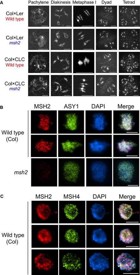
Figure 6. MSH2 accumulates on meiotic chromatin during early prophase I.

- Representative DAPI‐stained spreads of pachytene, diakinesis, metaphase I, dyad and tetrad meiotic stages, for wild type and msh2, in Col × Ler or Col × CLC hybrid backgrounds.
- Male meiocytes immunostained for MSH2 (red) and ASY1 (green), and stained for DAPI (blue) in wild type (Col) or msh2.
- As for B, but immunostaining wild type male meiocytes for MSH2 (red), MSH4 (green) and staining chromatin with DAPI (blue).
