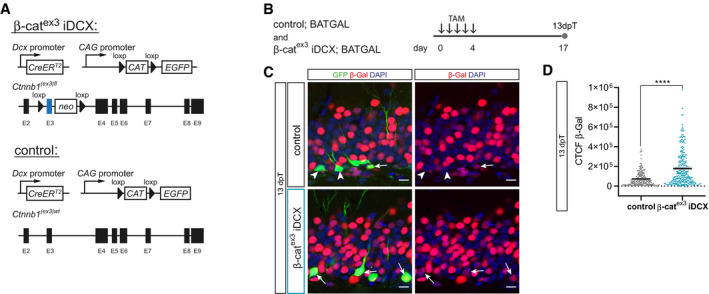
Schematic representation of the conditional alleles and transgenes of the β‐catex3 iDCX mouse line and control iDCX mice.
To analyze the impact of β‐catenin stabilization on canonical Wnt signaling, β‐catex3 iDCX mice and controls were crossed with BATGAL mice. Tamoxifen (TAM) was applied i.p. every 12 h for 5 days. Animals were analyzed 13 days post‐tamoxifen (dpT).
Representative images of β‐galactosidase reporter expression (red) in GFP‐positive cells (green) in control iDCX BATGAL and β‐catex3 iDCX BATGAL mice. DAPI in blue. Arrows and arrowheads indicate β‐galactosidase reporter‐positive and negative cells, respectively. Note the prominent β‐Gal signal intensity of surrounding non‐recombined cells in the granule cell layer of the DG. Scale bar = 10 μm.
Measurements of corrected total cell fluorescence (CTCF) of the β‐Gal signal in recombined cells at 13 dpT showed an increase in canonical Wnt signaling activity upon stabilization of β‐catenin in β‐catex3 iDCX BATGAL mice (P < 0.0001; control BATGAL: n = 205 cells from three animals, β‐catex3 iDCX BATGAL: n = 248 cells from three animals).
Data information: Data represented as mean ± SEM, significance was determined using two‐tailed Mann–Whitney
U‐test, and significance levels are displayed in GP style (****
P < 0.0001).