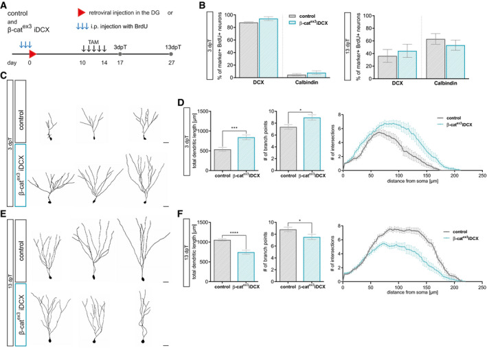
Experimental scheme for birthdating of adult‐born neurons and recombination. For morphology analysis, CAG‐RFP was stereotactically injected into the DG (C–F), for marker expression analysis, BrdU was injected i.p. every 24 h for 3 days (B). Tamoxifen was applied i.p. every 12 h for 5 days from day 10 to day 14. Mice were sacrificed 3 days post‐tamoxifen (dpT) and 13 dpT, which corresponds to 17 and 27 dpi, respectively.
Expression of DCX and Calbindin in BrdU+ neurons was comparable between experimental groups at 3 and 13 dpT (control: n = 5 animals, β‐catex3 iDCX: n = 5 animals).
Representative reconstructions of control and β‐catex3 iDCX neurons expressing RFP at 3 dpT. Scale bar = 20 μm.
Quantification of dendritic length (P = 0.0003), branch points (P = 0.0433), and Sholl analysis (P < 0.0001) showed a higher dendritic complexity of β‐catex3 iDCX neurons at 3 dpT (control: n = 22 cells from four animals, β‐catex3 iDCX: n = 19 cells from five animals).
Representative reconstructions of control and β‐catex3 iDCX neurons expressing RFP at 13 dpT. Scale bar = 20 μm.
Quantification of dendritic length (P < 0.0001), branch points (P = 0.0495), and Sholl analysis (P < 0.0001) showed a lower dendritic complexity of β‐catex3 iDCX neurons at 13 dpT (control: n = 20 cells from four animals, β‐catex3 iDCX: n = 19 cells from five animals).
Data information: Data represented as mean ± SEM, significance was determined using two‐way ANOVA for Sholl analysis and two‐tailed Mann–Whitney
‐test for all other analyses, and significance levels are displayed in GP style (*
< 0.0001).

