
Figure 5. Canonical Wnt signaling activity is decreasing with increasing age.
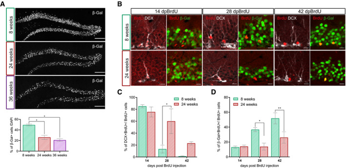
- Representative overview images and quantification of the β‐galactosidase reporter signal in the DG of 8‐week-old, 24-week‐old, and 36-week‐old BATGAL mice. Note the decrease in reporter signal with increasing age (n = 4 animals per age, 200–300 cells per animal; 8 weeks vs. 24 weeks P = 0.0268, 8 weeks vs. 36 weeks P = 0.0268). Scale bar = 100 μm.
- Representative images of BrdU+ cells (red) expressing DCX (gray) or β‐galactosidase (green) at 14 days post‐BrdU injection (dpBrdU), 28 and 42 dpBrdu in 8‐week-old and 24-week‐old BATGAL mice. Scale bar = 10 μm.
- Percentage of BrdU+ cells expressing DCX in 8‐week-old and 24-week‐old BATGAL mice (14 dpi: n = 3 animals, 28 dpi: n = 3 animals, 42 dpi: n = 3 animals). Note the prolonged expression of DCX in 24‐week-old mice (28 dpBrdU P = 0.0155).
- Percentage of BrdU+ cells expressing the β‐galactosidase reporter in 8‐week-old and 24-week‐old BATGAL mice (14 dpi: n = 3 animals, 28 dpi: n = 3 animals, 42 dpi: n = 3 animals). Note the reduced reporter expression in 24‐week-old mice (28 dpBrdU P = 0.0117, 42 dpBrdU P = 0.0034).