-
A
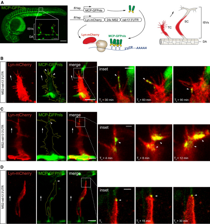
Left: Tg(fli1ep:MCP‐GFPnls) zebrafish embryo at 26 h post‐fertilisation (hpf) displaying vascular‐specific expression of MCP‐GFPnls. Inset shows the nuclear expression of MCP‐GFPnls in the intersomitic vessels (ISVs) sprouting from the dorsal aorta (DA). Middle: scheme depicts the in vivo MS2 system strategy with fli1 enhancer/promoter (fli1ep)‐driven expression of reporter constructs, simultaneous translation of Lyn‐mCherry reporter and binding of MCP‐GFPnls to 24xMS2‐rab13 3′UTR. Right: scheme illustrates ISV cells expressing Lyn‐mCherry imaged in panels B–D. TC: tip cell; SC: stalk cell.
-
B–D
Time‐lapse microscopy of Tg(fli1ep:MCP‐GFPnls) tip and stalk cells displaying mosaic expression of Lyn‐mCherry‐24xMS2‐rab13 3′UTR in ISV cells.
Data information:
T
0 = 24 hpf (C), 28 hpf (B), 48 hpf (D). Arrowheads indicate non‐nuclear localisation of MCP‐GFPnls; arrows indicate direction of ISV sprouting; yellow dashed lines outline ISV (A) or ISV cell (B‐D) borders; scale bars = 200 μm (A), 20 μm (B, D) and 10 μm (C); scale bars in insets = 20 μm (A), 5 μm (B, D) and 2 μm (C).

