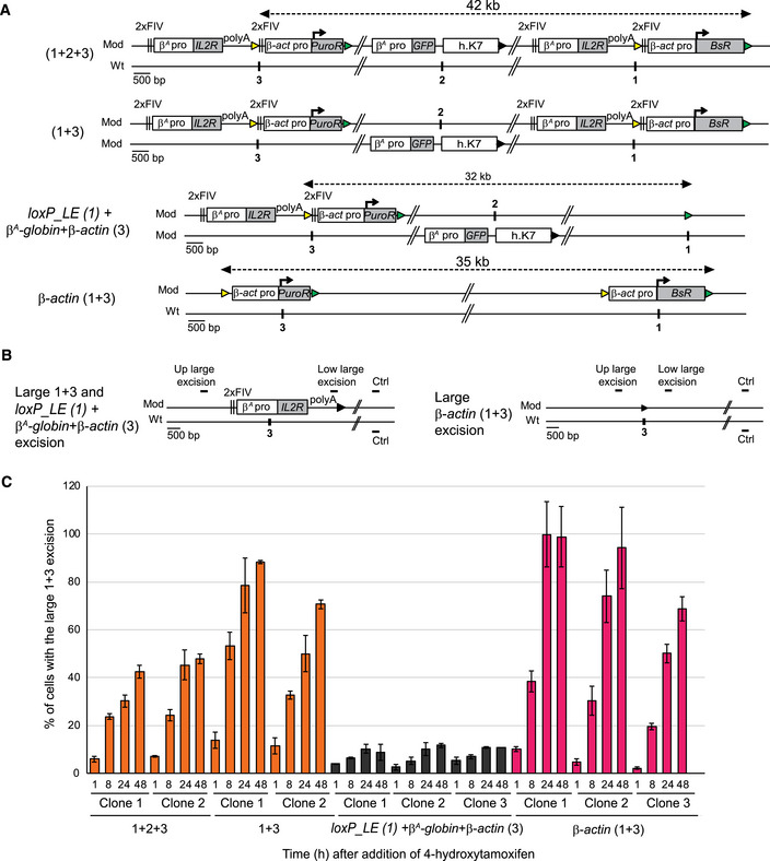
Figure 5. The formation of the early‐replicating domain is associated with the establishment of local interactions between the two advanced replicons separated by 30 kb.

- Clones 1 + 2 + 3, 1 + 3, and β‐actin (1 + 3) described in Fig 3, together with control clones containing one β A ‐globin + β‐actin construct at site 3 and one loxP_LE sequence inserted at site 1 on the same chromosome with the GFP reporter construct inserted at site 2 on the other chromosome loxP_LE (1) + β A ‐globin + β‐actin (3), were tested for their capacity to recombine after induction of the Cre recombinase. Yellow and green triangles represent reactive loxP_RE and loxP_LE sites, respectively, and black triangles represent recombined inactive loxP sites. Black vertical bars represent insertion sites.
- Recombination between the upstream loxP_RE element (yellow triangle) inserted at site 3 and the downstream loxP_LE element (green triangle) inserted at site 1 leads to a large 1 + 3 and loxP_LE (1) + β A ‐globin+β‐actin (3) or β‐actin (1 + 3) excision product.
- After 1, 8, 24, and 48 h of 4‐hydroxytamoxifen treatment, genomic DNA was extracted and quantified by semi‐quantitative PCR (see Appendix Fig S6). Error bars correspond to the standard deviation for PCR duplicates. The percentages of cells with the large 1 + 3 and loxP_LE (1) + β A ‐globin + β‐actin (3) or β‐actin (1 + 3) excision at each time point are shown for different cell lines.
