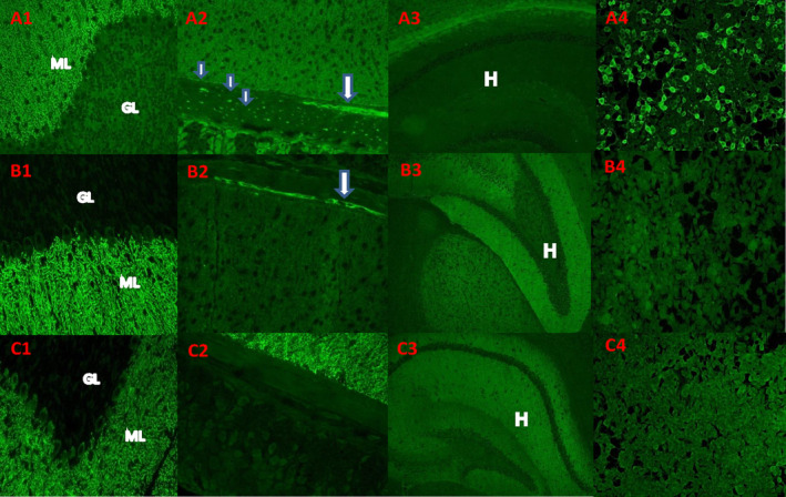
FIG. 1.

Examples of “medusa‐head” immunostaining pattern and IgG reactivity with ARHGAP26 (GRAF)‐transfected cells. (A) IgG in serum of patient 5 bound to neural elements in fixed cryosectioned mouse tissues. (A1) Staining is prominent in the molecular layer (ML) of the cerebellar cortex in purkinje neuronal dendritic arbors and (A2) in the gastric smooth muscle myenteric ganglia (arrow) and nerve fibers (small arrow). (A3) Hippocampus was not immunoreactive. (A4) human embryonic kidney 293 (HEK293) cells transfected with ARHGAP26/GRAF were highly reactive with patient's serum IgG; interspersed nontransfected cells were not stained. (B, C) The medusa head–like pattern of staining observed in 104 cases lacking ARHGAP26‐IgG specificity and excluded from this report: IgG in 2 such patients' sera yielded a medusa‐head pattern of staining that shares some similarities with ARHGAP26‐IgG, but does not bind to HEK293 cells transfected with ARHGAP26/GRAF (B4, C4). Unlike ARHGAP26‐IgG, these patients' serum IgG stains hippocampus brightly (B3, C3). Patient C serum does not stain myenteric ganglia or nerve fibers in the stomach smooth muscle (C2).
