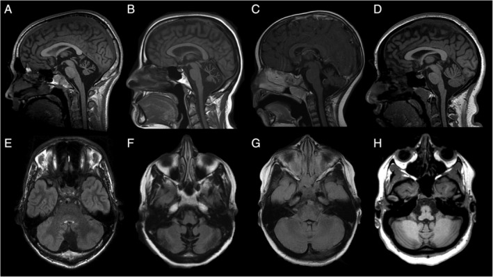
FIG 2.

Neuroimaging characteristics of 4 patients diagnosed with channelopathies. Brain magnetic resonance imaging of patients 620 (A,E; CACNA1A), 671 (B,F; CACNA1G), 423 (C,G; ITPR1), and 614 (D,H; KCNC1). Sagittal T1 (A–D) and axial fluid‐attenuated inversion recovery (E–H) images show atrophy of the cerebellar vermis and hemispheres.
