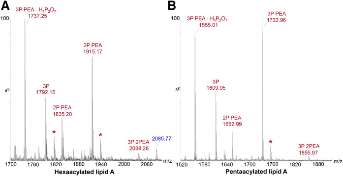
Fig. 4.
Negative-ion MALDI-TOF (non-IMS) spectra of intact LOS from N. meningitidis strain 90/90 (A) and strain 399 (B), which is a lpxL1 mutant strain, showing regions containing peaks for lipid A fragment ions produced by in-source decay. In the spectrum of 90/90 LOS (A) peaks for hexaacylated lipid A fragment ions with three P and two PEA groups can be observed at m/z 2038.26. Analogous peaks with three P and two PEA groups can be observed in the spectrum of 399 LOS (B) for penta-acylated lipid A that is lacking a laurate group on the nonreducing terminal glucosamine at m/z 1,855.97. More prominent peaks can be observed for fragment ions of lipid A with either two or three Ps and a single PEA moiety and three Ps. The prominent peaks at m/z 1,737.25 (A) and 1,555.01 (B) are most likely due to the facile loss of H4P2O7 from fragment ions at m/z 1,915.17 and 1,732.96, respectively, for lipid A with three P substituents and one PEA substituent. Peaks denoted with asterisks represent sodiated fragment ions (M-H2+Na)−. The peak at m/z 2,085.77 labeled in blue-colored font (A) is from OS fragment ions.

