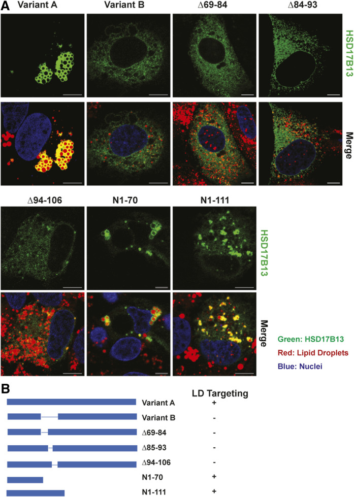
Fig. 2.
Identification of domains critical for LD targeting of HSD17B13. A: HepG2 cells were transiently transfected with HSD17B13 wild-type, the naturally occurring variant B (Δ71-106), or mutant plasmids and treated with fatty acids to induce LDs. Proteins were C-terminally tagged with GFP, which was used to determine their cellular localization (green). Nuclei were counterstained with Hoechst (blue), and LDs were stained with LipidTox (red). Images were analyzed by confocal microscopy. The bar indicates 10 μM. B: Schematic representation of full-length HSD17B13 (variant A) and mutant proteins. The plus sign indicates targeting to LDs; the minus sign indicates no targeting.

