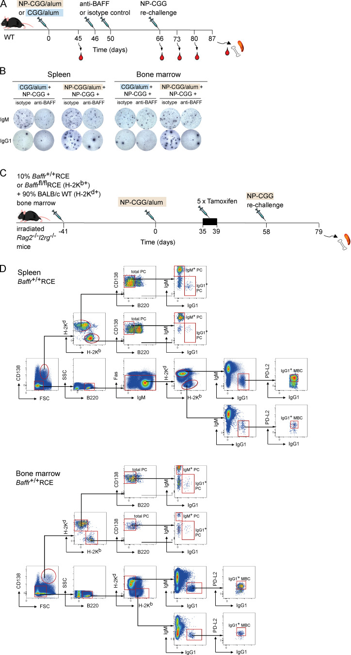
Figure S5.
Analysis of recall responses in mice treated with anti-BAFF or following deletion of BAFFR. (A) WT mice were immunized with CGG or NP-CGG in alum on day 0, followed by anti-BAFF or isotype control antibody injections 46 d and 50 d later and rechallenged with NP-CGG 16 d later. Blood was withdrawn 45, 66, 73, 80, and 87 d after primary immunization, and spleen and bone marrow were analyzed at day 87. (B) Images of example ELISPOT wells showing NP+ IgM+ or IgG1+ antibody secreting cells in spleen and bone marrow from mice treated as indicated. Each spot corresponds to a single antibody secreting cell. (C) Mixed bone marrow chimeras were generated by reconstituting irradiated Rag2−/−Il2rg−/− mice with 10% bone marrow from Baffr+/+RCE or Baffrfl/flRCE B6 mice (H-2Kb+) and 90% bone marrow from WT BALB/c mice (H-2Kd+) for 41 d. Mice were immunized with NP-CGG in alum on day 0, given five daily tamoxifen injections starting on day 35, and rechallenged with NP-CGG in PBS on day 58. Spleen and bone marrow were taken for analysis on day 79. (D) Flow cytometric analysis showing gating of H-2Kb+ or H-2Kd+ splenic IgG1+ MBCs (CD138−B220+Fas−PD-L2+) and bone marrow IgG1+ MBCs (CD138−B220+PD-L2+) as well as splenic and bone marrow IgM+ and IgG1+ PCs (FSChiCD138hiB220−). Red boxes indicate gates used to identify cell populations. FSC, forward scatter; SSC, side scatter.

