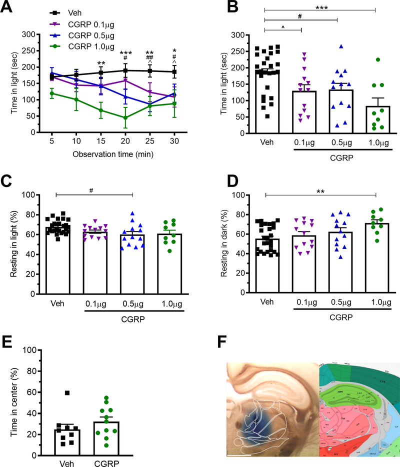
Figure 3. Injection of CGRP into the posterior thalamic region induces light-aversive behavior in dim light.
A. Time in light during 30 min light/dark assay following unilateral stereotactic injection of vehicle (Veh) or 0.1, 0.5, or 1.0 μg CGRP into the right posterior thalamic region of C57BL/6J mice. Light/dark testing was performed at 55 lux (dim light). All mice in panel A are further analyzed in panels B, C, D. B. Mean time in light per 5 min block of individual mice from panel A. C. Time resting in light during the assay. D. Time resting in dark during the assay. E. Time in center during the open field assay. F. Left: Site of injection of Evans blue dye. Right: Allen Mouse Brain Atlas coronal image representative of the injected area (image 74/132). Image credit: Allen Institute. Scale bar = 1000 μm. All error bars are SEM, * p < 0.05, ** p <0.01, *** P<0.001. # p < 0.05, ## p <0.01, ^ p <0.05. See Table 1 for detailed statistical analyses. A-D: Veh n=26, CGRP 1.0 μg n=9, 0.5 μg n=13, 0.1 μg n=13. E: Veh n=9, CGRP n=11.

