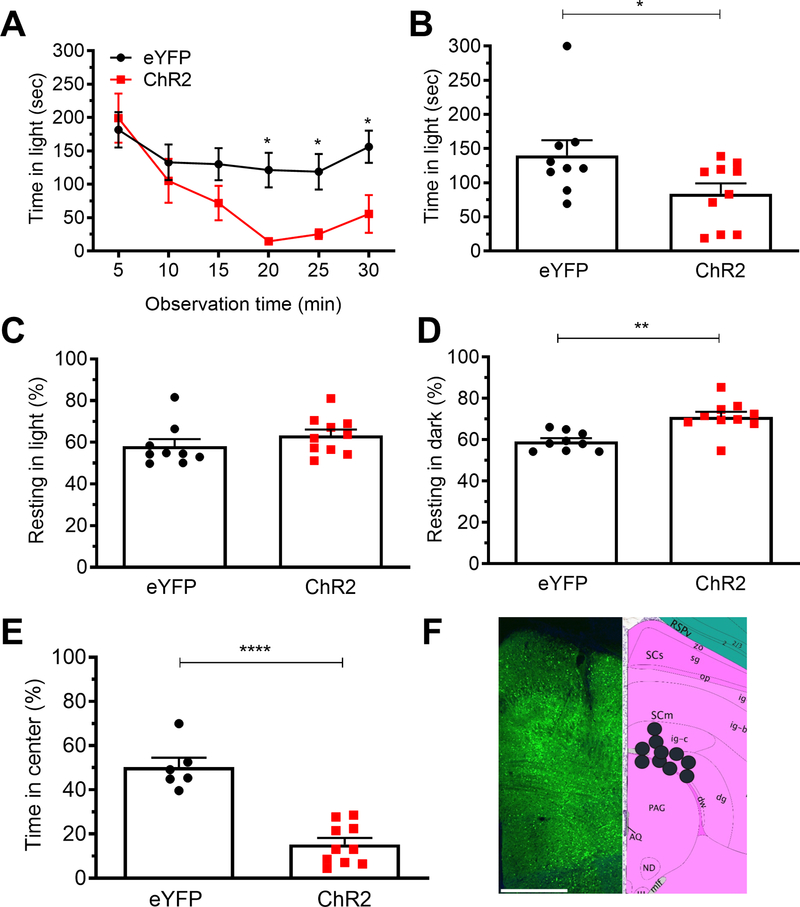
Figure 5. Optical stimulation of the dPAG induces light-aversive behavior accompanied by increased anxiety-like behavior.
A. Time in light over a 30 min light/dark assay in mice injected with AAV encoding either ChR2 or eYFP control in the dPAG. All mice in panel A are further analyzed in panels B, C, D. B. Mean time in light per 5 min block of individual mice from panel A. C. Time resting in light during the assay. D. Time resting dark during the assay. E. Time in center during the open field assay. F. Left: expression of AAV2-CaMKIIa-ChR2-eYFP in the dPAG. Right: schematic of positions of fiber optic probe tips for dPAG mice superimposed on Allen Mouse Brain Atlas coronal image (image 90/132). Image credit: Allen Institute. Scale bar = 500 μm. All error bars are SEM, * p < 0.05, ** p < 0.01, **** p < 0.001. See Table 1 for detailed statistical analyses. A-D: eYFP n=9, ChR2 n=10. E: eYFP n=6, ChR2 n=10.

