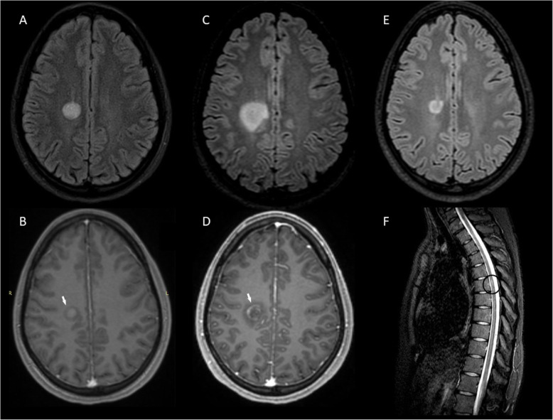
Fig. 4.
MRI of Case 7 before and after treatment with rituximab. A BCS-type lesion in the right centrum semiovale (A) with peripheral almost complete Gd + at outer layer (B arrow) at symptom onset. Follow-up MRI at 1 month, after corticosteroid treatment, showing enlargement of the BCS-type lesion (C) with Gd + (D arrow) and one lesion at Th6 level (F circle). BCS-type lesion (E) 4 months from the first rituximab course, 5 months from BCS onset. a, c, e: 3D FLAIR images. b, d: 3D T1-weighted contrast-enhanced images. f: STIR image. BCS: Baló’s concentric sclerosis, Gd+: gadolinium enhancement

