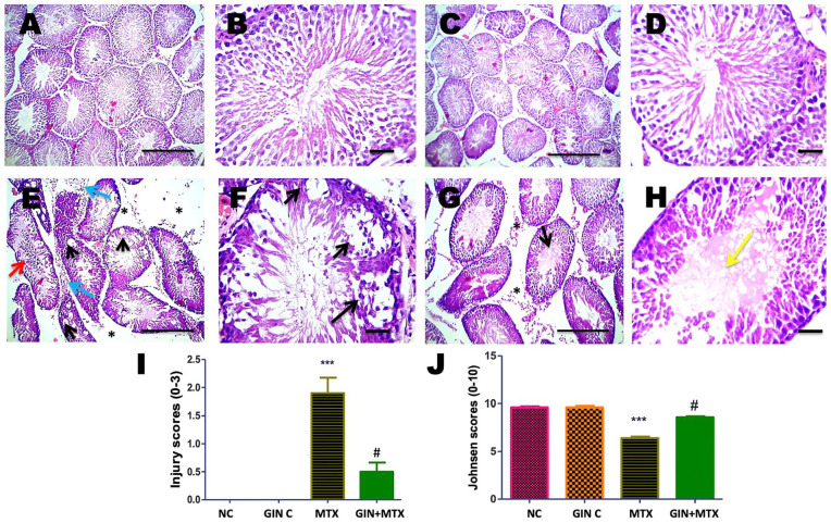
Figure 8.
Histopathological pictures of testicular tissues stained with hematoxylin and eosin (H and E) in all groups. Testis showed normal histopathology of seminiferous tubules with full spermatogenesis in NC (A and B) and GIN C (C and D) groups, interstitial edema (asterisks), leukocytic cells infiltration (blue arrows), marked vacuolization of spermatocytes (black arrows), desquamated spermatocytes (red arrow), necrotic spermatids and lowered spermatogenesis in MTX group (E and F). Interstitial edema (asterisks), partially restored spermatogenesis, hyalinization of spermatids is detected in lumen of few seminiferous tubules (yellow arrow) in GIN+MTX treated group (G and H). A, C, E, G X100, bar = 100 µm and B, D, F, H X 400, bar = 50 µm. Statistical analysis of testicular injury (I) and Johnsen (J) scores. ***P < .05 versus NC, #P < .05 versus MTX.

