Figure 5.

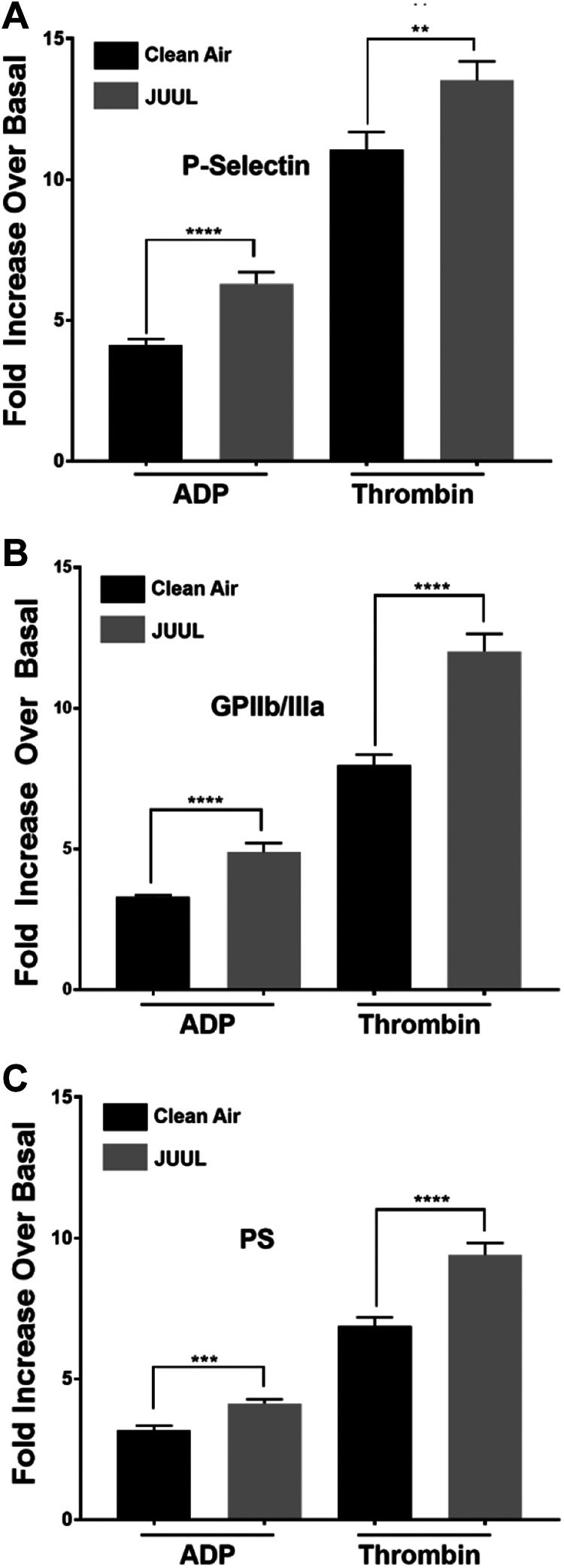
Platelets α granule secretion, integrin GPIIb/IIIa activation, and phosphatidylserine (PS) exposure are increased in JUUL-exposed mice. Washed platelets from clean air-exposed and JUUL e-cigarette-exposed mice were washed and activated with 1 μM ADP or 0.1 U/mL thrombin. Next, these platelets were incubated with FITC-conjugated CD62P antibody, before the fluorescence intensity was measured by flow cytometry (A). The average of the mean fluorescence intensities is shown (****P < .0001). Platelets were incubated with phycoerythrin-conjugated JON/A antibody, before the fluorescence intensity was measured by flow cytometry (B). The average of the mean fluorescence intensities is shown (****P < .0001). Platelets were incubated with fluorescein isothiocyanate–conjugated Annexin V antibody, before the fluorescence intensity was measured by flow cytometry (C). The average of the mean fluorescence intensities is shown (****P < .0001). These experiments were repeated 3 times, using blood that was pooled from 5 to 8 mice each time. Data are presented as mean ± SD.
