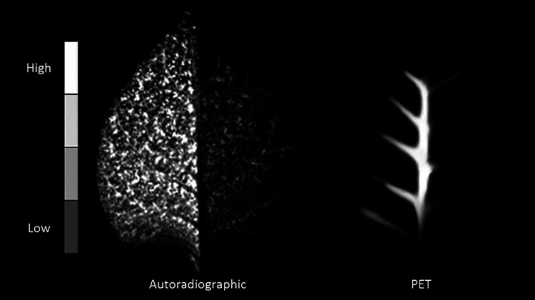
Figure 1.
Leaf uptake and distribution of [18F]F− imaged by autoradiography and PET. The images differ because autoradiography (left) detects escaped positrons (from decays that occur within the positron’s range of the plate), but because those escaped positrons annihilate outside the thinner, non-vascular leaf tissues they are not imaged by PET (right). The PET image, however, preferentially highlights tracer abundance within vascular tissue where positron annihilation occurs before positrons escape from these thicker structures. In both cases of imaging, the right side of an intact tobacco leaf from different plants is shaded causing more tracer to transport to the left-side.

