. 2020 Oct 1;2(5):e200389. doi: 10.1148/ryct.2020200389

Figure 2:

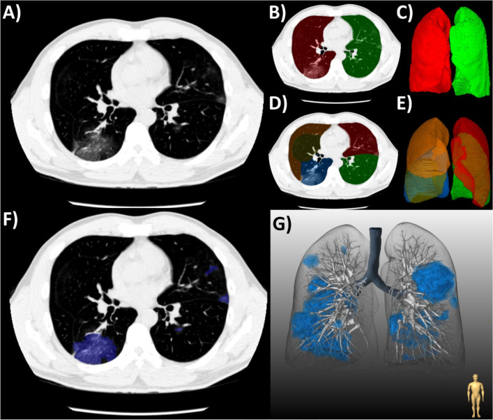
(a) Workflow for the quantification of COVID-19 pneumonia on chest CT in a patient with typical ground-glass opacities. (b-c) First, both lungs and (d-e) their respective lobes were automatically segmented by a deep-learning algorithm. Second, semi-automated segmentation of lesions was performed using (f) axial slices and shown with (g) three-dimensional rendering.

(a) Workflow for the quantification of COVID-19 pneumonia on chest CT in a patient with typical ground-glass opacities. (b-c) First, both lungs and (d-e) their respective lobes were automatically segmented by a deep-learning algorithm. Second, semi-automated segmentation of lesions was performed using (f) axial slices and shown with (g) three-dimensional rendering.