. 2020 Oct 1;2(5):e200389. doi: 10.1148/ryct.2020200389

Figure 5.

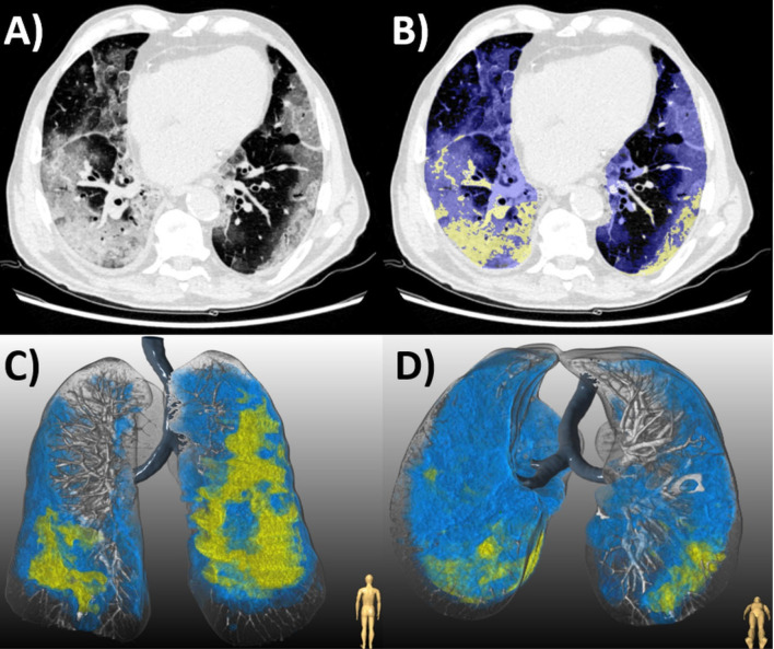
(a) Chest CT of an 87-year-old man with COVID-19 pneumonia who died 10 days later. (b) Axial slice shows bilateral diffuse ground glass opacities (GGO, blue) and consolidation (yellow). Lesion quantification revealed a GGO burden of 44.0% and consolidation burden of 8.0%. Three-dimensional renderings depict the distribution of disease in (c) coronal and (d) axial planes.

(a) Chest CT of an 87-year-old man with COVID-19 pneumonia who died 10 days later. (b) Axial slice shows bilateral diffuse ground glass opacities (GGO, blue) and consolidation (yellow). Lesion quantification revealed a GGO burden of 44.0% and consolidation burden of 8.0%. Three-dimensional renderings depict the distribution of disease in (c) coronal and (d) axial planes.