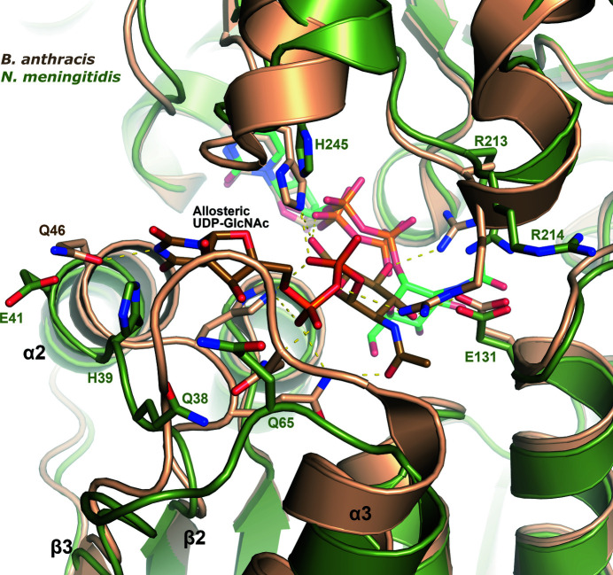
Figure 6.
Allosteric UDP-GlcNAc binding site. A superposition is shown of NmSacA (green) on UDP-GlcNAc 2-epimerase from B. anthracis (PDB entry 3beo; sand), the structure of which was determined with UDP-GlcNAc bound in the allosteric site (brown C atoms) and UDP bound in the active site. Shown are side chains that interact with the allosteric UDP-GlcNAc in the B. anthracis structure, with the corresponding NmSacA residues labeled in green. Yellow dashed lines show interactions with allosteric UDP-GlcNAc in B. anthracis. All residues are conserved in binding UDP-GlcNAc except for Glu41, which is Gln in the B. anthracis epimerase. The UDP-GlcNAc bound in the NmSacA active site is also shown (light green C atoms).

