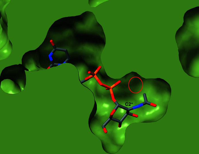
Figure 7.
Slice through a space-filling representation of the NmSacA structure with UDP-GlcNAc bound in the active site. The protein is shown as a green surface with the UDP-GlcNAc shown in stick representation with gray C atoms. The C2′′ atom is labeled along with the small pocket near the N-acetyl group (red circle). The structure explains how NmSacA can accept small modifications of the N-acetyl methyl group in UDP-GlcNAc analogs.

