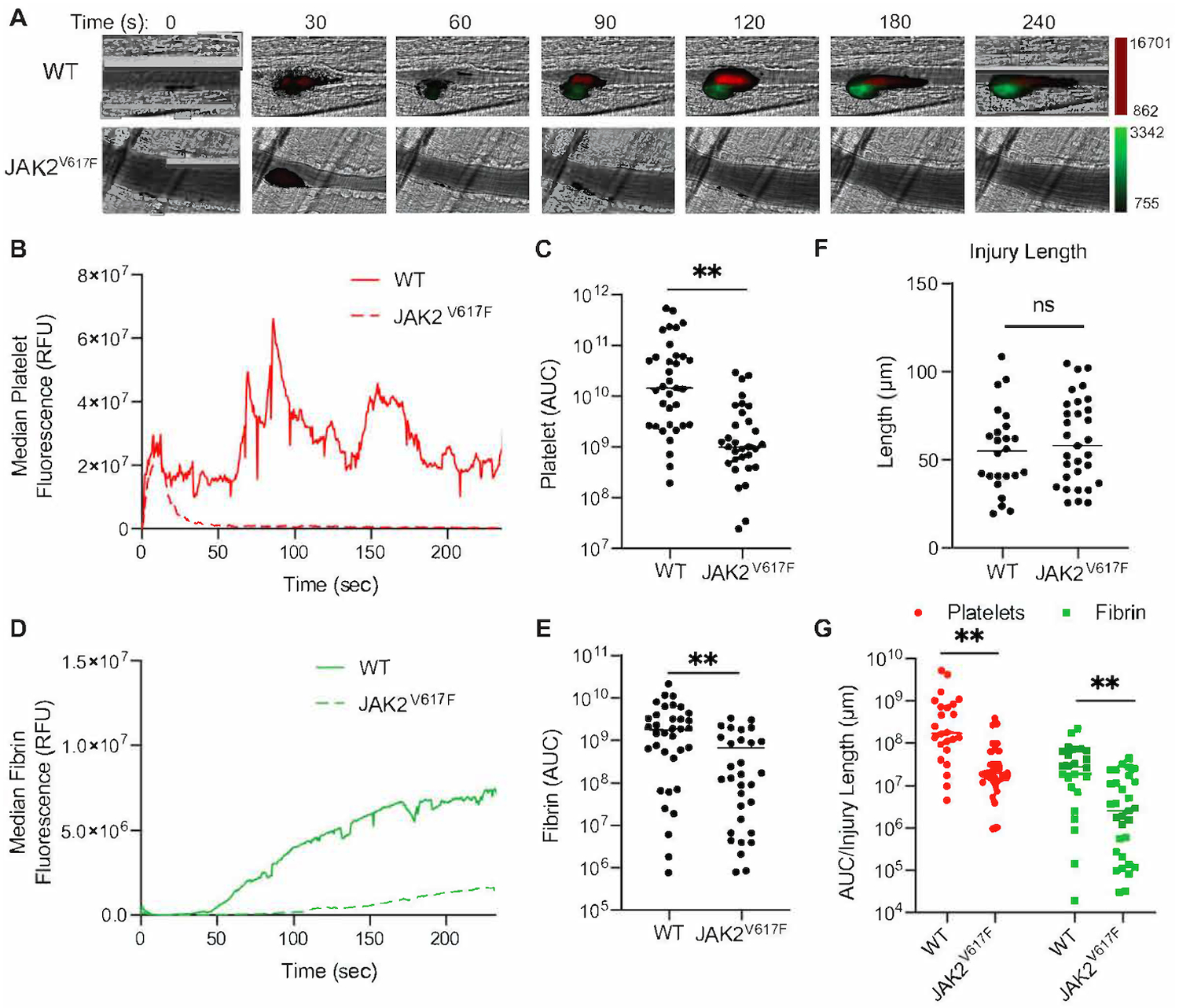
Figure 2.

JAK2V617F mice demonstrate decreased platelet accumulation and fibrin formation. (A) 28.4 weeks old males (n=5 each) WT and JAK2V617F mice were injected with Dylight 647-labeled anti-platelet antibody (CD42b) and Dylight 488-labeled anti-fibrin antibody (59D8). Representative images obtained at the indicated times following laser-induced injury are shown. Fluorescence intensity in each channel was normalized to background fluorescence obtained from images prior to laser-induced injury. (B) Median integrated platelet intensities following laser injury were calculated and plotted at 0.5 sec intervals for all thrombi in WT (n=35 thrombi) and Vav1-hJAK2V617F mice (n=31 thrombi). (C) Platelet AUCs were calculated and plotted for each thrombus formed in WT and Vav1-hJAK2V617F mice. Median values are indicated. (D) Median integrated fibrin intensities following laser injury were calculated and plotted at 0.5 sec intervals for all thrombi in vehicle and Vav1-hJAK2V617F mice. (E) Fibrin AUCs were calculated and plotted for each thrombus formed in WT and Vav1-hJAK2V617F mice. Median values are indicated. (F) Ablation injury lengths were measured in WT and Vav1-hJAK2V617F mice as a marker of injury severity. Median values were plotted. (G) Platelet accumulation and fibrin formation data were normalized for injury severity by dividing the AUC value for each thrombus by its corresponding ablation injury length. ns: not significant, **p < 0.01, Mann Whitney U test.
