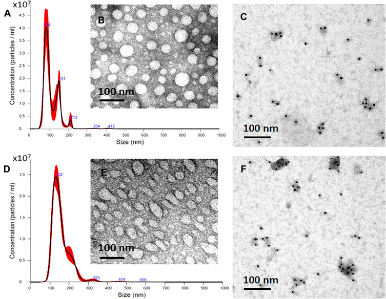
Figure 2.
Characterization of GMs-derived exosomes. Representative images of the size distribution of exosomes, as analyzed by NTA (A, D), The morphology and shape of exosomes, as analyzed by TEM (B, E), and CD63 positive dots of exosomes, as analyzed by immunogold EM (C, F). Exosomes isolated from SF7761 stem-cell like GMs (A–C) and U251 (D–F) GMs, respectively, were used in the experiment.

