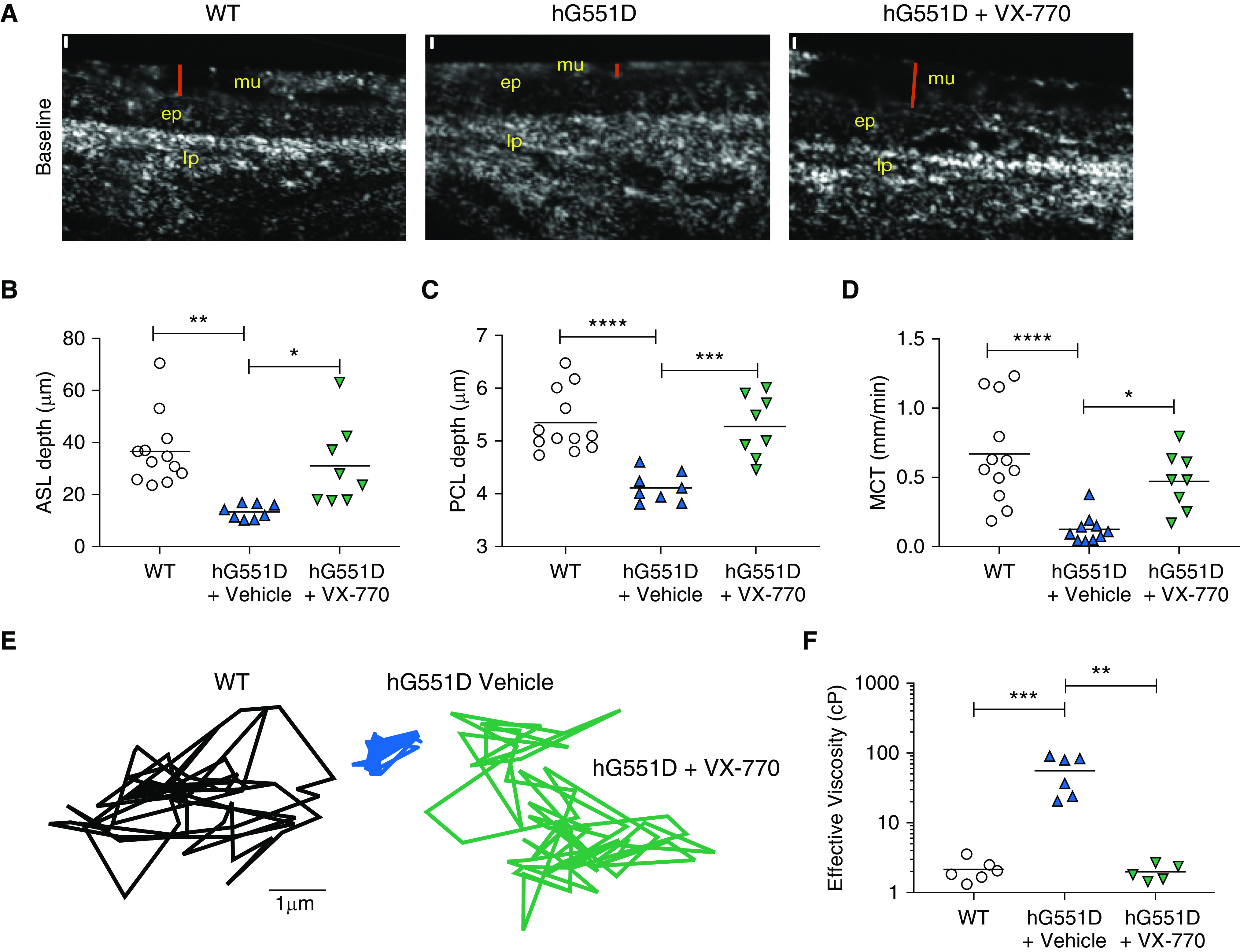
Figure 5.

The hG551D airway defect is rescued by VX-770 administration. Tracheae from 3-month-old wild-type and hG551D rats were imaged with μOCT. (A) Representative images of wild-type rats, hG551D rats, and hG551D rats treated with VX-770 depict the surface analyzed (red bar = airway surface liquid). Scale bars (white), 10 μm. Quantitative analysis of the images yields (B) airway surface liquid depth, (C) periciliary layer depth, and (D) mucociliary transport rates. After the ciliary beating was arrested, particles were added to the surface and Brownian motion was tracked. (E) Representative tracings depict freedom of movement in the mucus from each trachea. (F) Conversion of mean squared displacement yielded effective viscosities. *P < 0.05, **P < 0.01, ***P < 0.001, and ****P < 0.0001. ASL = airway surface liquid; ep = epithelial layer; lp = lamina propria layer; MCT = mucociliary transport; mu = mucus layer; PCL = periciliary layer; WT = wild-type.
