Figure 1.

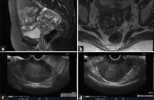
Image study of the pelvic myoma. Pelvic magnetic resonance image showing sagittal (a) and coronary (b) views of the myoma. Pelvic ultrasound showing sagittal (c) and coronary (d) views of the myoma

Image study of the pelvic myoma. Pelvic magnetic resonance image showing sagittal (a) and coronary (b) views of the myoma. Pelvic ultrasound showing sagittal (c) and coronary (d) views of the myoma