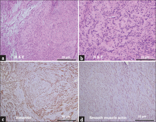
Figure 3.

Pathology of pelvic myoma. (a and b) Histology of the myoma showed by H and E staining. Smooth muscle cells were elongated with eosinophilic cytoplasm. Smooth muscle bundle showed fascicular pattern. The immunohistochemistry was positive for vimentin (c) and smooth muscle actin (d) which are typical of myoma. Scale bar = 10 μm (b) and = 20 μm (a, c, and d)
