Figure 2.
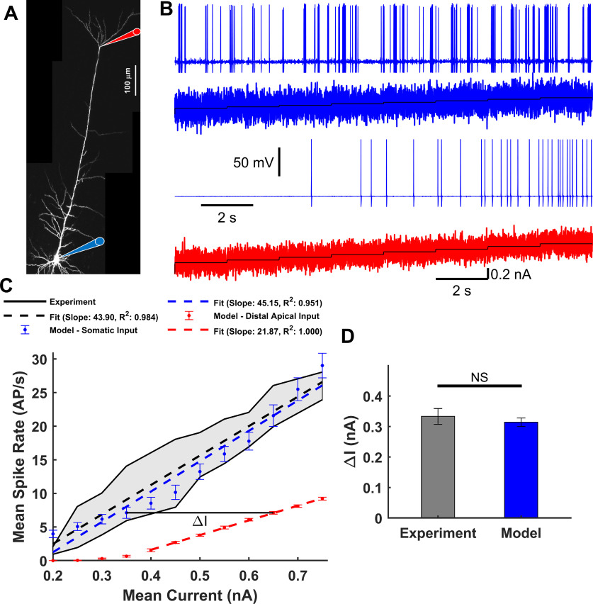
f-I relationship. A, Micrograph of an L5-PC with recording locations at the soma (blue) and distal trunk (red) indicated with diagram pipettes. B, Somatic Na+-AP responses (first and third panels) to the staircase incremented noisy input current (second and fourth panels) injected into the soma (blue) and distal trunk (red). C, Observed (black) (Larkum et al., 2004; Bahl et al., 2012) Na+-AP firing frequency as a function of the mean input current. Gray fill represents the range of observed values. Simulated mean and SEM spike rate over 50 trials for each current step in the soma (blue) or distal trunk (red) compartment. Superimposed are observed (black dashed) and simulated (blue and red dashed) linear regressions. D, Current differences (ΔI) between the f-I curves for somatic and distal trunk stimulation to produce the same Na+-AP firing frequency. No significant differences were found between the observed ΔI, numerically estimated from Larkum et al. (2004) and that predicted by the model (t(5) = 2.0789, p = 0.0922, paired two-tailed t test).

