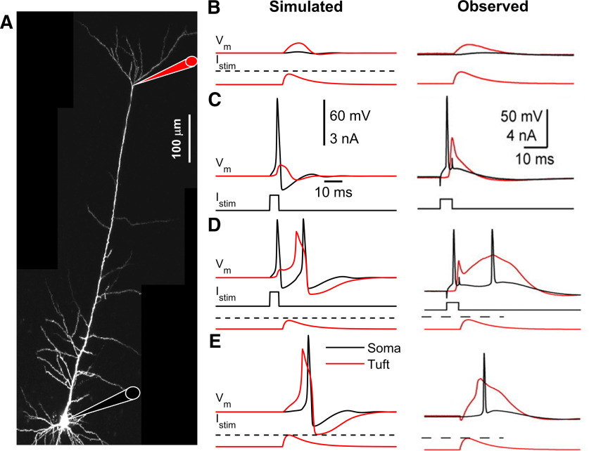
Figure 3.
Back-propagating AP-activated Ca2+ spike firing. A, Micrograph of a L5-PC with recording locations at the soma (black) and distal trunk (red) indicated with a schematic pipette. B, Simulated (left) and observed (right) (Schaefer et al., 2003) subthreshold current injected into the apical dendrites creates only subthreshold somatic and dendritic depolarization. C, Simulated and observed suprathreshold somatic current pulse elicits an Na+-AP that propagates back to the apical dendrites creating a dendritic depolarization but no dendritic Ca2+ spike. D, Simulated and observed combined somatic and tuft stimulation evokes an Na+-AP, a dendritic Ca2+ spike, and another somatic Na+-AP following the onset of the dendritic Ca2+ spike. E, Simulated and observed suprathreshold stimulation of distal apical dendrites evokes a dendritic Ca2+ spike. C, Scales are common for all simulated (left) and observed (right) results. Red represents apical-dendrites/trunk. Black represents basal-dendrites/soma. Dashed line indicates dendritic threshold. For visualization, the ∼10 mV shift in membrane resting potential from the somatic to the apical-dendrites/trunk compartment was removed.

