Figure 4.
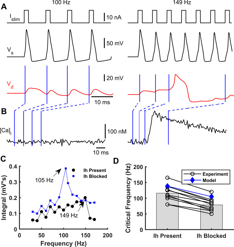
Effect of somatic stimulation frequency on dendritic Ca2+ spike occurrence. A, A simulated train of brief suprathreshold pulses at frequencies of 100 Hz (left) and 149 Hz (right) (top) was injected into soma eliciting a train of Na+-APs (black, below). Only the 149 Hz train evoked a dendritic Ca2+ spike (red, bottom). B, Intracellular dendritic Ca2+ concentration during somatic stimulation at 100 Hz (left) and 149 Hz (right), respectively. Blue lines indicate the Ca2+ concentration at each time instant of the dendritic voltage traces indicated in A. C, Integrated area below the dendritic voltage traces as a function of the Na+-AP frequency with (blue) and without (black) the Ih current. CFs of 105 Hz and 149 Hz were obtained when Ih was present and absent, respectively. D, Observed shift in CF after blocking Ih for 11 cells (black circles, numerically estimated from Berger et al., 2003) and simulated with the model (blue diamonds).

