Figure 6.
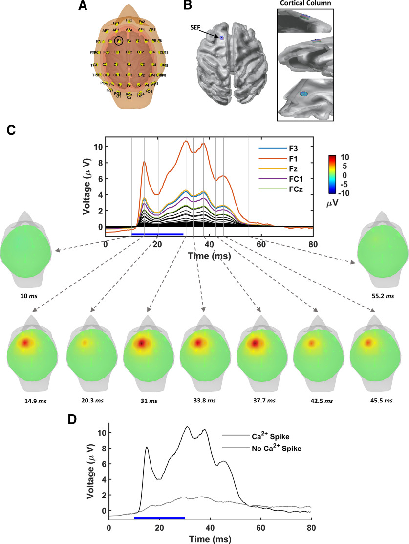
Large-scale EEG signatures of subcellular dendritic Ca2+ spikes generated by a collection of L5-PCs located in SEF. A, Monkey's head model with EEG electrodes positioned according to the international 10-10 system. Tissue compartments are indicated in different colors. B, Location of the simulated functional cortical column in the SEF. Right, Expanded view of the functional cortical column from different perspectives. C, Top, EEG potentials at each electrode resulting from brief supra-CF stimulation (blue horizontal bar) of the collection of L5-PCs simulating optogenetic stimulation in Suzuki and Larkum (2017). Scalp potentials at each electrode contact are averaged over 10 simulated trials, each randomly affected by system noises in the membrane potentials and the calcium concentrations. Bottom, Topographical voltage maps illustrated for indicated times (gray vertical attached to dashed arrows). D, EEG potential sampled by electrode F1 (circled A), which is the closest electrode to the functional cortical column. Samples are shown with (black) and without (gray) discernible Ca2+ spikes.

