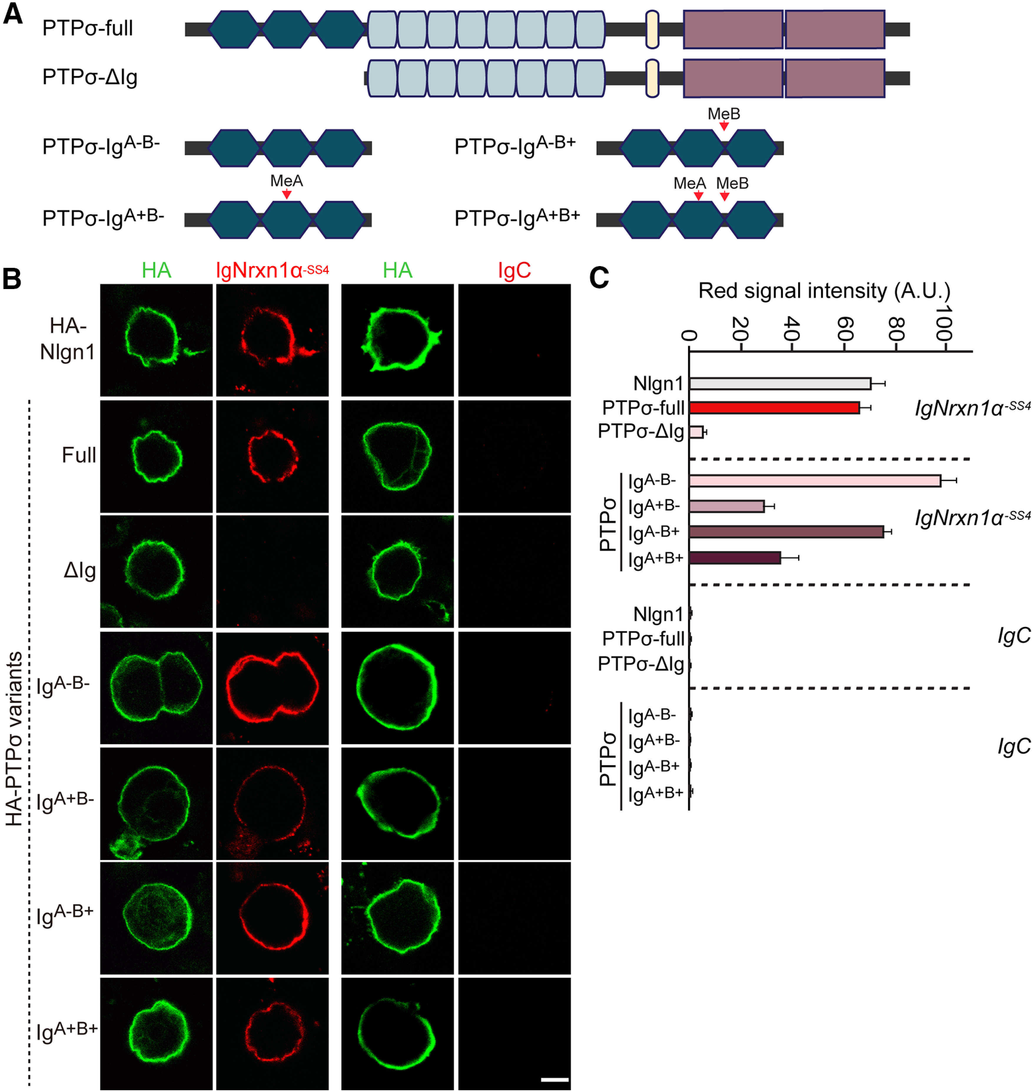
Figure 7.

The PTPσ Ig domain is necessary and sufficient for interaction with Nrxn1α. A, Schematic diagrams of PTPσ WT and deletion mutants. B, Representative images of cell-surface binding assays. HEK293T cells expressing HA-Nlgn1, HA-PTPσ WT, HA-PTPσ ΔIg, or the indicated PTPσ Ig domain splicing variants constructs were incubated with 10 μg/ml of control IgC or Ig-Nrxn1α-SS4 and then analyzed by immunofluorescence imaging of Ig-fusion proteins (red) and HA antibodies (green). Scale bar, 10 μm. C, Quantification of the average red intensities in the green positive region of HEK293T cells in B. n indicates the number of cells as follows: Ig-Nrxn1α/Nlgn1, n = 27; Ig-Nrxn1α/PTPσ-full, n = 33; Ig-Nrxn1α/PTPσ ΔIg, n = 32; Ig-Nrxn1α/PTPσ IgA–B–, n = 30; Ig-Nrxn1α/PTPσ IgA+B–, n = 22; Ig-Nrxn1α/PTPσ IgA–B+, n = 27; Ig-Nrxn1α/PTPσ IgA+B+, n = 23; IgC/Nlgn1, n = 10; IgC/PTPσ-full, n = 11; IgC/PTPσ ΔIg, n = 15; IgC/PTPσ IgA–B–, n = 11; IgC/PTPσ IgA+B–, n = 11; IgC/PTPσ IgA–B+, n = 12; and IgC/PTPσ IgA+B+, n = 10.
