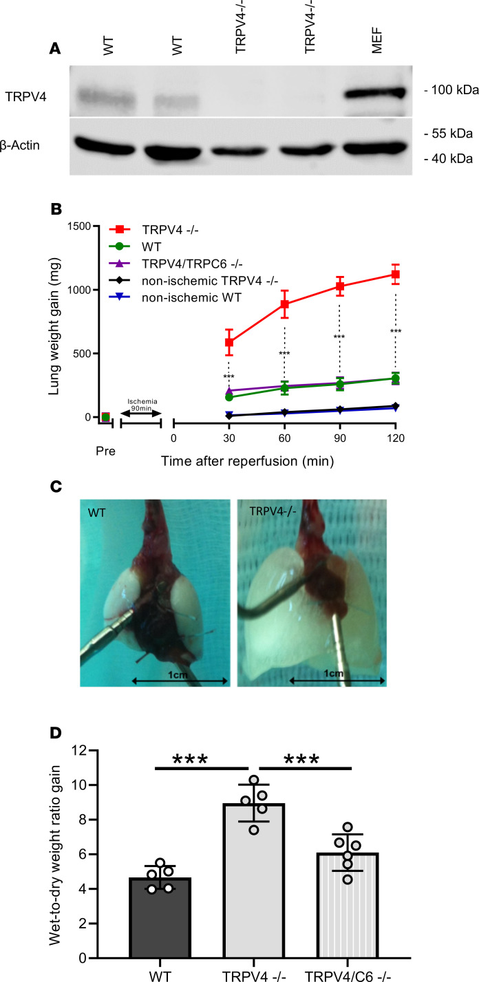
Figure 1. Ablation of TRPV4 increases ischemia-induced edema formation in mouse lungs.
(A) TRPV4 protein expression in mouse lungs was evaluated by immunoblotting in whole-lung lysates of WT and TRPV4-deficient (TRPV4−/−) mice using a TRPV4-specific antiserum. Murine embryonic fibroblasts (MEFs) served as an additional positive control. β-Actin was used as loading control. (B) Constant weight measurement of ischemic and nonischemic WT and TRPV4–/– and TRPV4/TRPC6 double-deficient (TRPV4/TRPC6–/–) isolated perfused lungs. (C) Representative images of WT and TRPV4–/– lungs after ischemia. (D) Wet-to-dry weight ratio gains of TRPV4–/– and TRPV4/TRPC6–/– lungs compared with those of WT controls. Data represent mean ± SEM of at least 5 lungs for each genotype. Significance between means was analyzed using ANOVA (B and D); ***P < 0.001.

