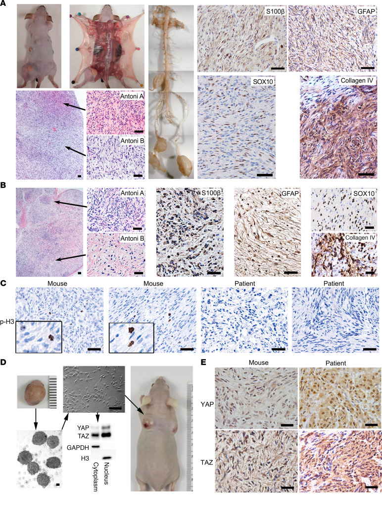
Figure 1. Hippo pathway inactivation in Hoxb7+ lineage cells results in multiple schwannoma formation.
(A) Dissection and histological characterization of mouse schwannoma: H&E and IHC of Schwann cell markers (S100β and GFAP), a neural crest marker (Sox10), and collagen IV. (B) H&E and IHC of S100β, GFAP, SOX10, and Collagen IV on human schwannoma tissue sections. (C) IHC of phospho-Histone H3 on mice and human schwannoma tissue sections. (D) Mouse schwannomas were harvested, and tumorsphere cell culture was performed. Tumorspheres were then seeded to fibronectin-coated plates for monolayer culture. Both cytoplasmic and nuclear fractions isolated from these monolayer cultured cells were analyzed by Western blotting. The tumor cells were injected into nude mice s.c. (n = 6). (E) IHC of YAP and TAZ on mouse and human schwannoma tissue sections. Scale bars: 50 μm.

