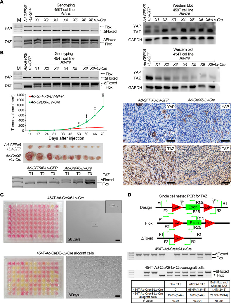
Figure 5. Canonical Hippo signaling through YAP/TAZ is required for schwannomagenesis.
(A) Genotyping (left) and Western blot (right) of YAP and TAZ in 459T cells serially infected with Ad-GFP/Lv-GFP or Ad-Cre/Lv-Cre. (B) Genotyping (left top panel) and Western blot (right top panel) of YAP and TAZ in 454T cells serially infected with Ad-GFP or Ad-Cre/Lv-Cre; tumor volume of 454T-Ad-GFPX6-Lv-GFP and 454T-Ad-CreX6-Lv-Cre schwannoma tumor in nude mice (middle left panel) (n = 10/group); gross picture of tumors from experimental endpoint (lower left panel) (n = 10/group); IHC (lower right panel) and genotyping analysis of TAZ (lower left panel) in transplanted nude mice tumor tissue and its derived tumor cell lines. (C) Single cell clonal analysis of 454T-Ad-Crex6-Lv-Cre cells (upper panel) and their transplanted tumor–derived cells (lower panel). Yellow cell culture media indicates the proliferation of single cell clone; pictures were taken at 28 days (454T-Ad-Crex6-Lv-Cre cells) and 8 days (transplanted tumor–derived cells). (D) Single cell nested PCR for TAZ. Diagram of PCR primer design for detecting the flox or Δfloxed allele of TAZ in single cell level. F1, R1, and R1.5 primers were used for the first PCR. F2, R2, and R2.5 primers were used for the second PCR (upper panel). Representative pictures of gel electrophoresis for single cell nested PCR products (middle panel). Quantification of flox and Δfloxed alleles of TAZ from 454T-Ad-Crex6-Lv-Cre cells and their allograft-derived cells (lower panel). 454T-Ad-Crex6-Lv-Cre cells, n = 45. 454T-Ad-Crex6-Lv-Cre allograft cell, n = 44. First lane, DNA marker; empty lane, failure to detect the signal. Each lane represents a single cell. Scale bars: 50 μm. Two-tailed Student’s t test was applied to evaluate statistical significance in B. Statistics are represented as the mean ± SEM. *P < 0.05, **P < 0.01.

