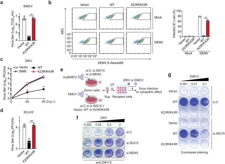
Figure 4. ISGylation is required for viral restriction by MDA5.
(a) EMCV titers in the supernatant of HEK293T cells that were transiently transfected for 40 h with either empty vector, or FLAG-tagged MDA5 WT or K23/K43R and then infected with EMCV (MOI 0.001) for 24 h, determined by TCID50 assay. (b) Percentage of DENV-infected MDA5 KO HEK293 cells that were transiently transfected for 24 h with either empty vector or FLAG-tagged MDA5 WT or K23R/K43R and then mock-treated or infected with DENV (MOI 5) for 48 h, assessed by FACS using an anti-flavivirus E (4G2) antibody. SSC, side scatter. (c) ZIKV titers in the supernatant of MDA5 KO SVGAs that were transiently transfected for 30 h with either empty vector, or FLAG-tagged MDA5 WT, K23R/K43R, or S88E and then infected with ZIKV (MOI 0.1) for the indicated times, determined by plaque assay. (d) SCoV2 titers in the supernatant of HEK293T-hACE2 cells that were transiently transfected for 24 h with either empty vector, or FLAG-tagged MDA5 WT or K23/K43R and then infected with SCoV2 (MOI 0.5) for 24 h, determined by plaque assay. (e) Schematic of the experimental approach to decouple the role of ISG15 in MDA5-mediated IFN induction from its role in dampening IFNAR signaling. (f) NHLF ‘donor’ cells were transfected for 40 h with the indicated siRNAs and then infected with mutEMCV (MOI 0.1) for 16 h. Cell culture supernatants were UV-inactivated and transferred onto Vero ‘recipient’ cells for 24 h, followed by infection of cells with ZIKV (MOI 0.002 to 2) for 72 h. ZIKV-positive cells were determined by immunostaining with anti-flavivirus E (4G2) antibody and visualized using the KPL TrueBlue peroxidase substrate. (g) RIG-I KO HEK293 ‘donor’ cells were transfected for 24 h with si.C or si.ISG15 and subsequently transfected with either empty vector or FLAG-tagged MDA5 WT or K23R/K43R for 24 h, followed by EMCV infection (MOI 0.001) for 16 h. UV-inactivated culture supernatants were transferred onto Vero ‘recipient’ cells for 24 h, followed by infection with EMCV (MOI 0.001 to 0.1) for 40 h. EMCV-induced cytopathic effects were visualized by Coomassie blue staining. Data are representative of at least two independent experiments (mean ± s.d. of n = 3 biological replicates in a, b, c). **p < 0.01 (unpaired Student’s t-test).

