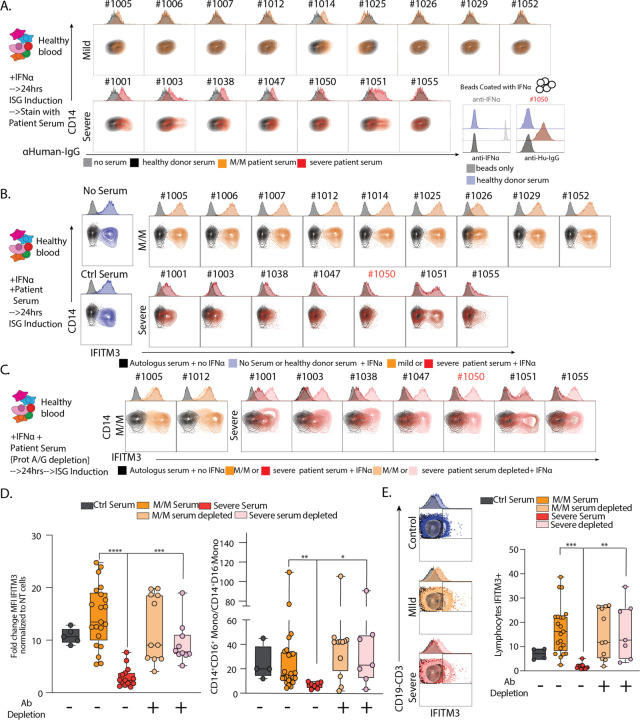
Figure 5: Neutralization of ISG induction by Antibodies from Severe COVID-19 Patients.
A. Contour plots and histograms of CD14 Monocytes from healthy blood cultured with IFNα to induce expression of ISGs and stained with serum from heathy donor, mild/moderate (M/M) or severe SARS-CoV-2 positive patients with secondary staining with α-human IgG. Bottom right: histogram of beads coated with IFNα and stained with an antibody raised against IFNα or serum from severe SARS-CoV-2 positive patient #1050 or healthy donor. Black histograms represent non coated beads. B-C. Contour plots and histograms of CD14 Monocytes from healthy blood cultured with IFNα and serum from heathy donor, mild/moderate or severe SARS-CoV-2 positive patient quantifying levels of intracellular IFITM3 staining. C. Mild/Moderate (light yellow) or Severe (pink) sera were pre-treated with protein G/A before incubation with PBMC. D. Box plot of IFITM3 induction in CD14 monocytes (left) and intermediate to classical monocytes ratio (right) from 2 different experiment and 2 different healthy donors. E. Left: Contour plots and histograms of pooled CD3+/CD19+ lymphocytes from healthy blood cultured with IFNα and serum from heathy donor, mild/moderate or severe SARS-CoV-2 positive patients. Mild/moderate (light yellow) or Severe (pink) sera were pre-treated with protein G/A before incubation with PBMC to deplete antibodies. Right: Box plot of IFITM3 induction in lymphocytes. Differences in D. and E. were calculated using a two-way ANOVA test with multiple comparisons. * p.value < 0.05; ** p.value < 0.01; *** p.value <0.001; **** p.value < 0.0001; ns: non-significant.

