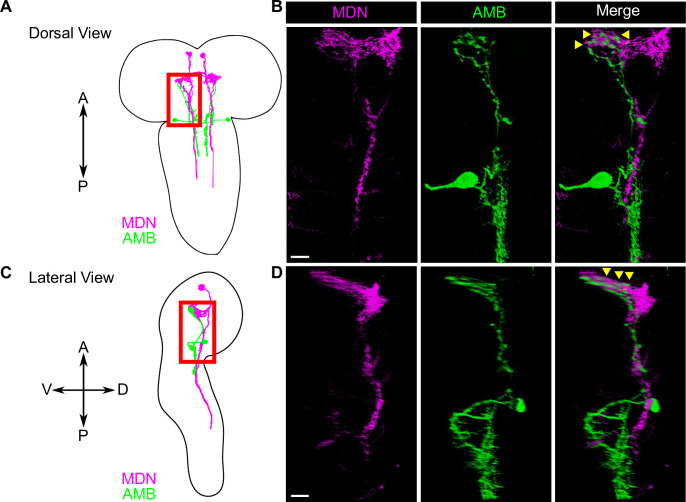
Fig 4. AMB axons are apposed to MDN dendrites in the larval brain.
(A-D) A schematic view of AMBs and MDNs from the dorsal (A) and from lateral side (C). The area with red boxes in (A) and (C) were shown in (B) and (D), respectively. Dual-labeling of AMBs (green) and MDNs (magenta). AMBs and MDNs were co-labeled with membrane-localized RFP and membrane-localized GFP, respectively. Genotypes: w; R60F09-LexA, tsh-GAL80/LexAop-rCD2RFP; R73F04-GAL4, Gad1-2A-GAL80/UAS-mCD8GFP. Scale bars, 10 μm.

