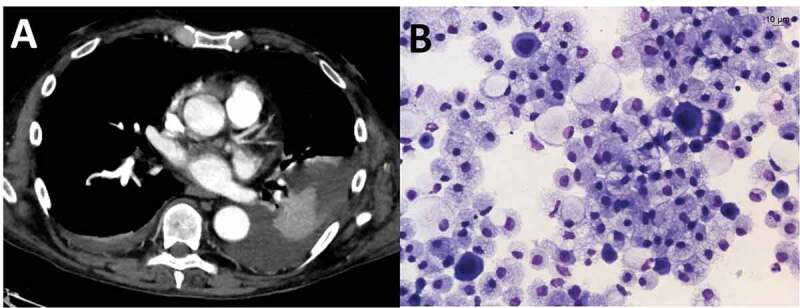
Figure 1.

(a): Chest CT after intravenous administration of only 60 ml of contrast medium. Virtual monoenergetic reconstruction (55 KeV) of dual-energy CT data shows pleural effusion and atelectasis of the neighboring lung parenchyma and excludes active foci of bleeding. (b): Microvacuolated macrophages and scattered mesothelial cells with enlarged multiple nuclei suggesting viral infection
