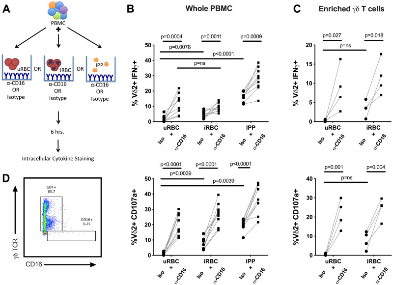
Fig 3. Stimulation through CD16 augments Vδ2 cell activation by P.falciparum.
(A) Schematic detailing stimulation conditions for data presented in panel B. (B) Percent of Vδ2 T cells producing IFNγ or positive for CD107a after stimulation of whole PBMC with uRBC, iRBC or IPP, with or without plate bound anti-CD16 crosslinking antibody (n = 9). (C). Percent of Vδ2 T cells producing IFNγ or positive for CD107a after stimulation of negatively selected γδ T cells with uRBC or iRBC, with or without plate bound anti-CD16 crosslinking antibody (n = 4). (D) Live cells positive for γδ TCR after negative selection. All Vδ2+ T cells shown in B-C were gated on singlets and CD3+ events to exclude NK cells. Samples for these experiments were selected from individuals with greater than 30% of their Vδ2 T cells expressing CD16. P values were determined by Wilcoxon matched pairs signed rank test.

